
Introduction
Diffuse idiopathic pulmonary neuroendocrine cell hyperplasia (DIPNECH) is a rare disorder characterized by the hyperplasia of pulmonary neuroendocrine cells.1,2 It classically presents as a chronic cough and dyspnea in women aged 50-60 years. Imaging typically reveals multifocal pulmonary nodules and mosaic attenuation.2 Given the non-specific symptomatology, DIPNECH may be initially misdiagnosed as asthma or COPD. Once formally diagnosed, DIPNECH may act as a confounder and lead to diagnostic delay of any secondary or co-existing pulmonary illness.2 Here, we present a case of DIPNECH treated with inhaled and systemic corticosteroids complicated by Mycobacterium abscessus Pulmonary Disease (M. abscessus PD) and tracheomediastinal fistula – co-existing diagnoses not yet described in the literature.
Case
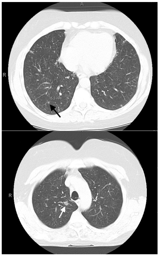
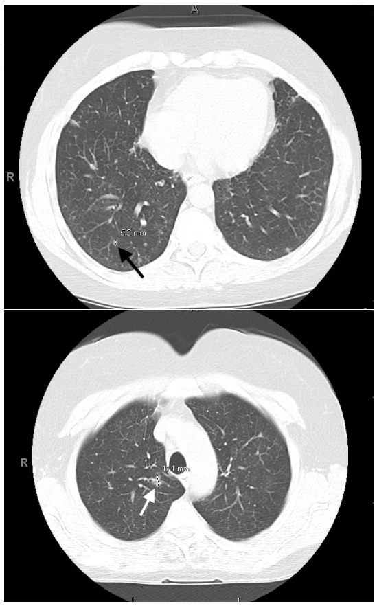
A 67-year-old female, a never-smoker with a history of childhood asthma, was referred to the pulmonary clinic for evaluation of progressive dyspnea, chronic cough, and innumerable small pulmonary nodules on CT scans spanning at least 7 years (Figure 1). She reported symptom progression over two years despite taking the equivalent of 3200 mg of inhaled fluticasone daily as well as several trials of 5-day-long oral prednisone courses. Pulmonary function tests (PFTs), including spirometry, diffusion capacity, and lung volumes, were normal. Positron emission tomography-computed tomography demonstrated an FDG-avid 8 mm right lower lobe nodule in the setting of diffuse background nodularity. Surgical lung biopsy (SLB) revealed carcinoid tumorlets and diffuse neuroendocrine cell hyperplasia, confirming DIPNECH. The patient was treated with routine high-dose inhaled corticosteroids (ICS) and cough suppressants. The patient declined somatostatin-analogue therapy due to personal preference.
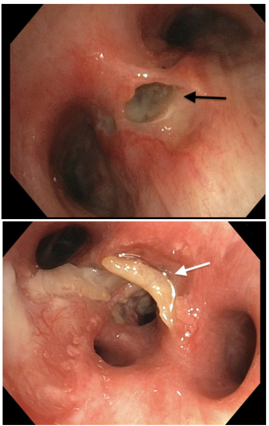
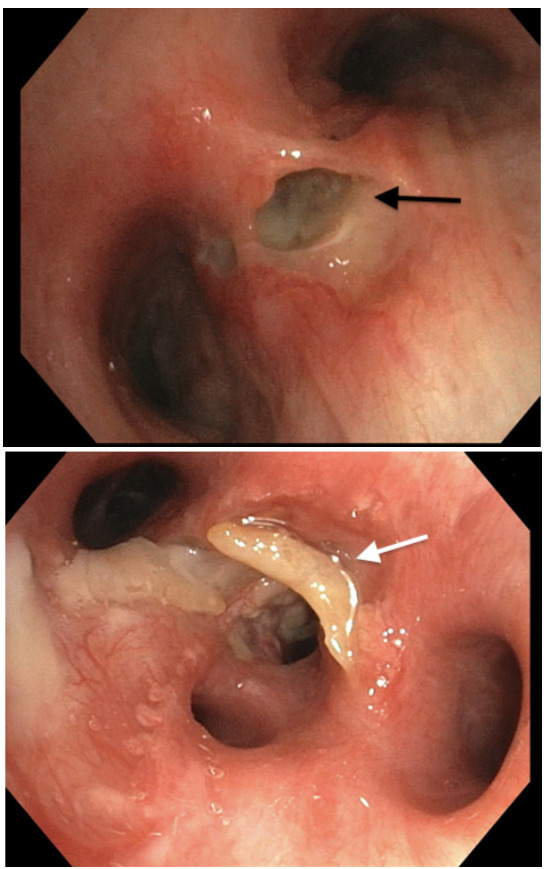
Despite treatment over the following year, the patient continued to have a persistent and debilitating cough. Repeat CT chest imaging revealed several new large pulmonary nodules and post-obstructive pneumonia. Subsequent bronchoscopy identified a necrotic tracheomediastinal fistula and an endobronchial lesion (Figure 2) that revealed M. abscessus on biopsy. Multidrug antimicrobial therapy was initiated but discontinued after 19 days due to severe adverse effects. Unfortunately, the patient continued to have severe symptomatology and ultimately transitioned to hospice care with medical aid in dying. She died one month after hospice admission.
Discussion
This case illustrates a classic presentation of DIPNECH, yet complicated by M. abscessus PD and a tracheomediastinal fistula. Despite suggestive imaging findings dating back seven years, DIPNECH remained undiagnosed until she was evaluated by pulmonology and underwent SLB. This delay in diagnosis was likely due to the low prevalence and awareness of DIPNECH in the medical community, as well as anchoring bias towards her history of asthma.2 After her DIPNECH diagnosis, this patient had a persistent cough and dyspnea. Ultimately, a CT Chest revealed new large nodules and an endobronchial lesion. This is highly atypical for DIPNECH alone, as DIPNECH usually follows an indolent course.1,2 DIPNECH can progress to metastatic carcinoid tumors, which can grow into the endobronchial space.2,3 In this case, however, bronchoscopy with biopsy revealed M. abscessus as the primary driver for her worsening symptomatology.
Literature exploring infectious complications of DPINECH primarily focuses on the importance of ruling out infectious etiologies for pulmonary nodularity during initial diagnosis, as reactive neuroendocrine hyperplasia can mimic DIPNECH.1,2,4 Few case reports discuss the interplay between Nontuberculous Mycobacteria (NTM) PD and DIPNECH, but all published literature features Mycobacterium Avium Complex (MAC) infection, and no published case reports highlight the co-diagnosis of DIPNECH and M. abscessus.5
Notably, NTM PD and DIPNECH share radiographic and symptomatic features, making the diagnosis of NTM PD in these patients uniquely challenging. Features of both diseases include diffuse small pulmonary nodules, cough, and dyspnea, though nodules in NTM PD patients are often associated with bronchiectasis.2,6 It is possible that patients with DIPNECH are at an increased risk of NTM PD, though further research is needed to understand any association. In this patient’s case, prolonged high-dose ICS exposure in addition to courses of systemic steroids was likely the primary risk factor for the development of NTM PD.
The patient’s tracheomediastinal fistula and M. abscessus endobronchial lesion are unique features of this case. NTM infections are rarely complicated by endobronchial lesions; previously reported cases occurred in immunocompromised patients, with only one prior case report highlighting an endobronchial lesion caused by M. abscessus.7
M. abscessus infection has been implicated in airway and pleural fistulization.8,9 Although cultures from this patient’s fistula site were negative, the clinical association with M. abscessus infection, along with visualized purulence, suggested this as the most likely etiology. Additionally, ICS exposure can cause tracheobronchial smooth muscle atrophy and may increase risk for fistula formation.10
In conclusion, this case highlights diagnostic challenges, potential infectious complications, and the need to reconsider prolonged use of high-dose ICS in suspected DIPNECH.