
Introduction
Primary mediastinal B-cell lymphoma (PMBL) is a non-Hodgkin B-cell lymphoma that originates from thymic B cells within the anterior mediastinum. While similar in presentation to diffuse large B-cell lymphoma (DLBCL), it is now recognized as a separate oncologic entity with specific molecular markers and treatment indications.1 PMBL and other B-cell lymphomas rarely present with cavitary pulmonary lesions. More likely etiologies in the differential for cavitary pulmonary lesions include infection (especially fungal), vasculitis, and certain malignancies.2 We describe a rare case of lymphoma initially presenting as a cavitary lung lesion, which was initially empirically treated as pulmonary coccidioidomycosis (due to positive IgG serologies and suggestive imaging findings), but was eventually correctly diagnosed as B-cell Lymphoma and later confirmed to be PMBL following genetic testing.
Case Report
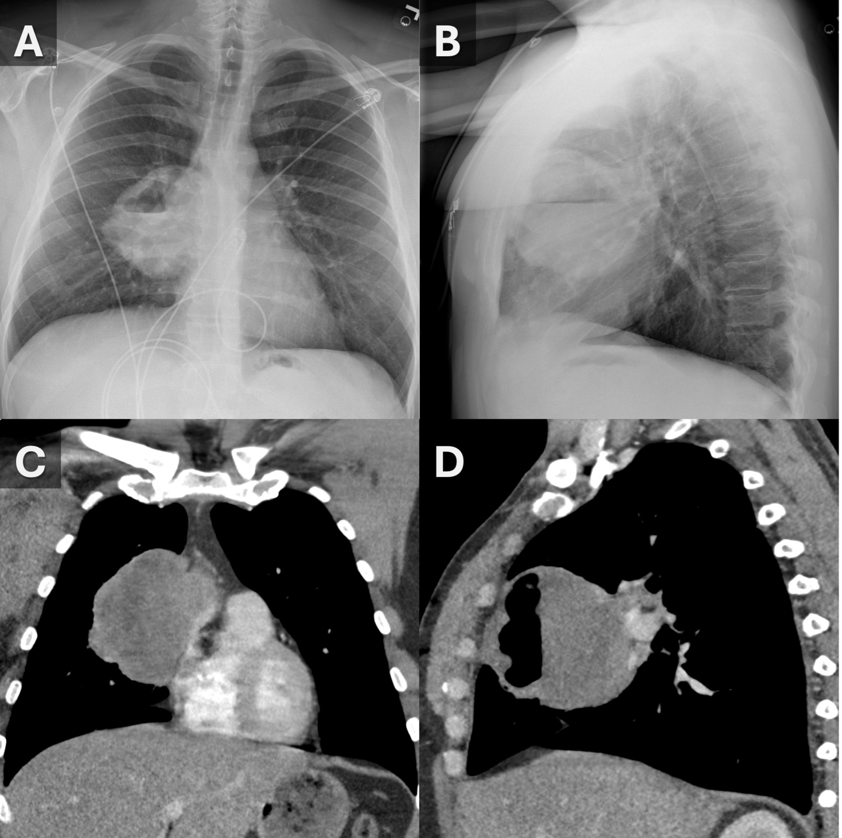
A 34-year-old man presented to an outside emergency department with a four-month history of worsening cough and right-sided pleuritic chest pain. No changes in weight, decrease in energy, or other B symptoms were reported. Baseline laboratory studies revealed: hemoglobin 14 g/dL, hematocrit 41%, WBC 6.5 x 10⁹/L (70% neutrophils, 20% lymphocytes, 3.5% eosinophils), and platelets 301 x 109/L. Initial chest X-ray (Figure 1A, B) revealed a large right-sided anterior mediastinal vs. paramediastinal pulmonary mass with an air-fluid level. This finding prompted a CT, which demonstrated a large, cavitary mass measuring 9 cm x 9.4 cm x 11 cm (Figure 1C, D). The mass appeared to be arising from the medial segment RML near the minor fissure and right heart border. Some additional satellite nodules, some with air bronchograms, were also noted in the right middle lobe. The patient was transferred to our institution for additional care.
_and_lateral_(b)_cxr_images_obtained_upon_initial_presentation_demonstrate_a_right_p.png)
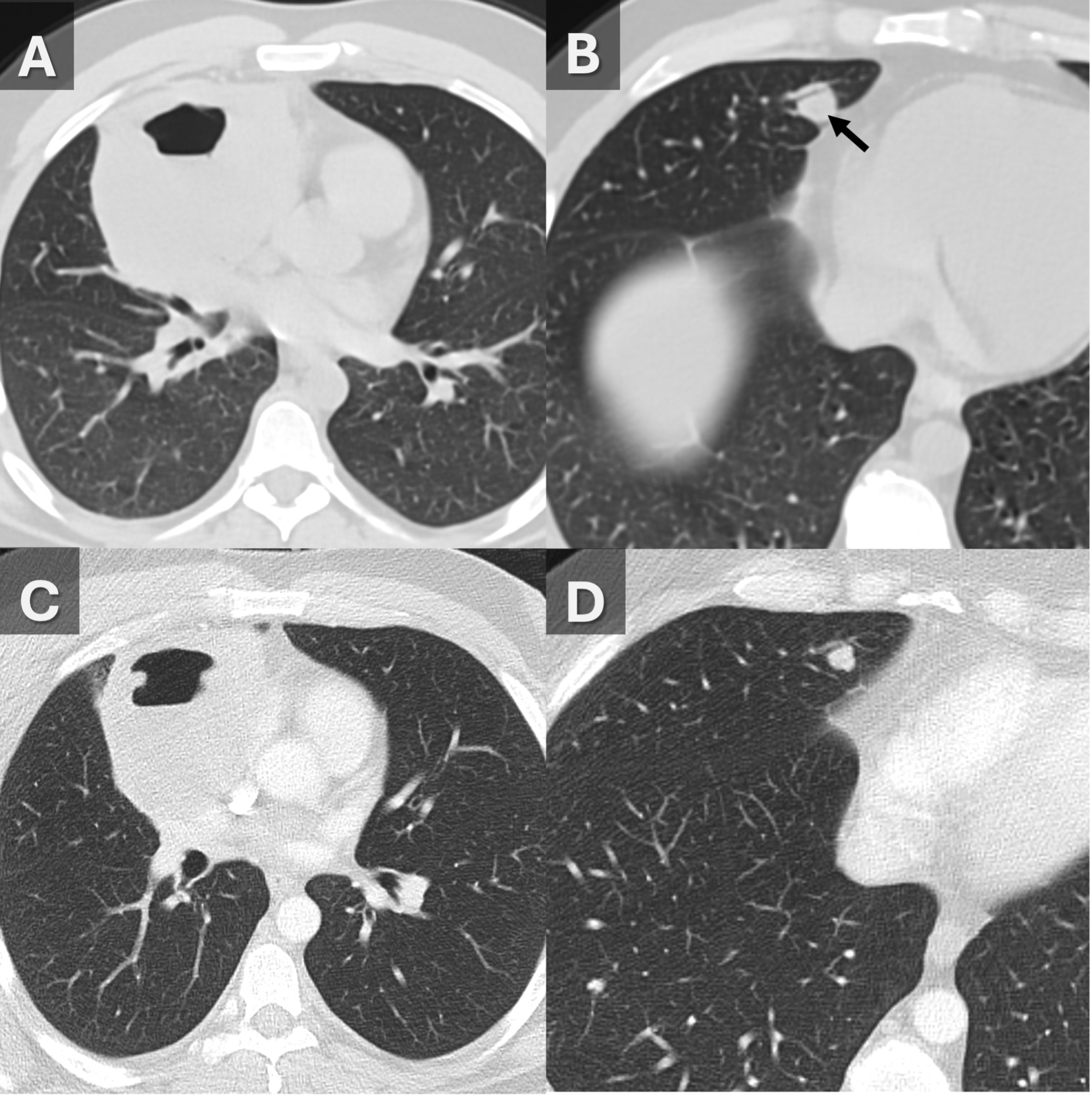
Infectious disease workup and testing for underlying immunodeficiencies at our institution were negative, except for positive coccidioidomycosis IgG serologies. Of note, the negative IgM serology seems somewhat discordant given the apparent large burden of infection based on imaging findings. FNA and BAL were performed utilizing robotic bronchoscopy. BAL cell count/differential was neutrophils 34%, lymphocytes 16%, alveolar macrophages 49%, and eosinophils 1%. The obtained specimen was non-diagnostic, however, the obtained cells tested negative for malignancy. The patient was empirically treated with fluconazole and scheduled for a follow-up CT in 1 month. The follow-up CT (Figure 2) demonstrated an interval increase in the mass and in the right middle lobe nodules despite a month of antifungal therapy. This discordant behavior prompted repeat tissue sampling via bronchoscopy, with a larger number of samples collected.
_.png)
Upon repeat bronchoscopy, a diagnosis of DLBCL was made. The following week, the patient was re-admitted to our institution with dyspnea, chest pain, cough, fevers, and neutrophil predominant leukocytosis, consistent with SIRS criteria. A repeat BAL noted positive rhinovirus/enterovirus infection. He was initiated on antibiotic coverage for community acquired pneumonia and started R-CHOP chemotherapy.
Subsequent analysis of the biopsy sample found the mass to be consistent with PMBL with CD30+ and CD23 partially positive, distinguishing the tumor from DLBCL. His treatment was transitioned to DA-REPOCH accordingly with acyclovir and TMP/SMX being added for prophylaxis. A follow-up 17-FDG Positron Emission Tomography-CT (PET-CT) performed after 2 weeks and again after another month demonstrated a rapid and substantial decrease in the size of the mass and the adjacent right middle lobe nodules.
Discussion
Cavitary lung lesions have a broad differential diagnosis, including infection, vasculitis, and malignancy. Although lymphomas very rarely cavitate prior to treatment, an important first step is to distinguish pulmonary from mediastinal origin to narrow the differential. Pulmonary masses are commonly associated with pyogenic lung abscesses, tuberculosis, pulmonary infarction, and malignancies.3 Mediastinal masses can also be caused by etiologies such as tuberculosis, fungal infections such as coccidioidomycosis and histoplasmosis, and neoplasms, including lymphoma.4
Internal gas can provide a clue of the lesion origin. Cavitary lesions of pulmonary origin often contain air, while those of mediastinal origin will invariably be filled with fluid/necrotic debris unless they have been biopsied, formed a fistula, gained access to atmospheric gas, or are infected with very rare gas-forming organisms. In the vast majority of cases where there is a question of mediastinal vs. pulmonary origin for a cavitary lesion, if the cavity is filled with gas, the lesion will usually be pulmonary in origin.
In addition to identifying cavity contents, imaging provides key features that help differentiate pulmonary from mediastinal origin. Lesions forming acute angles with the mediastinum suggest pulmonary origin, whereas lesions with obtuse angles are often mediastinal in origin.5 On chest X-ray, the hilum overlay sign is another tool that provides insight into tissue origin.5 Preserved hilar borders seen through a mass suggest it does not arise from the hilum, supporting a prevascular or paramediastinal location.5 CT imaging of pulmonary lesions often show spiculated or ragged margins, vascular convergence, pleural retraction, or ground-glass opacities in adjacent lung parenchyma while lesions of mediastinal origin are often smooth or lobulated and confined within mediastinal compartments.6 Our imaging (Figure 1) shows obtuse angles with mediastinum, a positive hilum overlay sign, and confinement to the prevascular mediastinal compartment, supporting a mediastinal origin. Non-Hodgkin lymphoma can present with secondary pulmonary involvement (Figure 2), which can demonstrate air bronchograms within consolidation.7
Another barrier to diagnosing lymphomas is the difficulty obtaining a biopsy. Excisional biopsies are usually not possible due to the location, and FNA biopsies often yield only fibrotic material, resulting in non-diagnostic specimens.8 In this patient, a FNA and BAL were performed in the original clinical encounter that both gave negative malignancy results. While the procedural technique remained unchanged, a repeat FNA with additional sample collections was required to acquire sufficient biopsy material. Prompt diagnosis and treatment are essential to reduce symptoms of continued growth, such as shortness of breath and chest pain.
DLBCL can have similar morphologies/presentations as PMBL and is often the initial diagnosis for PMBL, as seen in this case. However, PMBL is recognized as a distinct lymphoma with unique clinical behavior and molecular profiles.1 PMBL and DLBCL have different prognostic outcomes and treatments, with a 5-year OS of 85% and 60-70%, respectively.9 One treatment traditionally used for both lymphomas was R-CHOP combined with radiation therapy, but radiation therapy can be contraindicated in young patients, such as the one in this case, due to long-term cardiovascular toxicity and possible tissue damage.10 Another treatment, the dose-adjusted rituximab, etoposide, prednisone, vincristine, cyclophosphamide, and doxorubicin (DA-R-EPOCH) regimen, is an alternative with higher treatment rates in patients with PMBL (5-year OS rate 97%) and offers no radiation exposure, compared to R-CHOP (5-year OS rate 82%).10
Cavitary lung lesions are not typical presentations of lymphomas. While the prevalence of cavitary lymphoma in the lungs is not described in literature, they can often present with dyspnea and shortness of breath due to their size. Infectious causes may be initially suspected, but lymphomas and other malignancies should be ruled out. Important considerations in these cases are careful biopsy sample techniques aimed towards minimizing the chances of acquiring a non-diagnostic specimen. Accurate diagnosis through immunophenotypic confirmation of malignancies can guide specific treatment and maximize chances of positive patient outcomes.