
INTRODUCTION
Common variable immunodeficiency (CVID) comprises a heterogeneous group of disorders characterized by hypogammaglobulinemia and reduced antigen-specific antibody responses, resulting in impaired responses to vaccination, recurrent infections, and increased risk of autoimmune and lymphoproliferative complications. Pulmonary complications are a hallmark of CVID, affecting up to 85% of patients, and commonly manifest as recurrent lower respiratory tract infections, bronchiectasis, and interstitial lung disease, contributing significantly to morbidity and reduced quality of life.1 While infections such as sinusitis and gastrointestinal infections are well recognized, non-infectious manifestations—including enteropathy, lymphocytic colitis, interstitial lung disease, and lymphoma—are also clinically important.2,3
Granulomatous-Lymphocytic Interstitial Lung Disease (GLILD) occurs in approximately 8–20% of patients with CVID.4 According to the British Lung Foundation and the UK Primary Immunodeficiency Network, GLILD is defined as a distinct clinico-radio-pathologic ILD in patients with CVID, characterized by a lymphocytic infiltrate and/or granulomas in the lung, in whom other potential conditions have been considered and excluded.5 The condition usually manifests between 20 and 50 years of age, and females are more frequently affected. In approximately 15% of patients, GLILD is detected incidentally on imaging without overt symptoms. Clinically, patients most commonly present with cough and exertional dyspnea.6
Sarcoidosis is a common mimic of CVID-associated interstitial lung disease (CVID-ILD). Differentiation requires high-resolution computed tomography (HRCT) to assess distribution, morphology, and systemic features, as well as biopsy to confirm histopathology.
We present a case of GLILD manifesting as steroid-dependent ILD in a young adult, initially misdiagnosed as sarcoidosis, highlighting key diagnostic pitfalls.
CASE
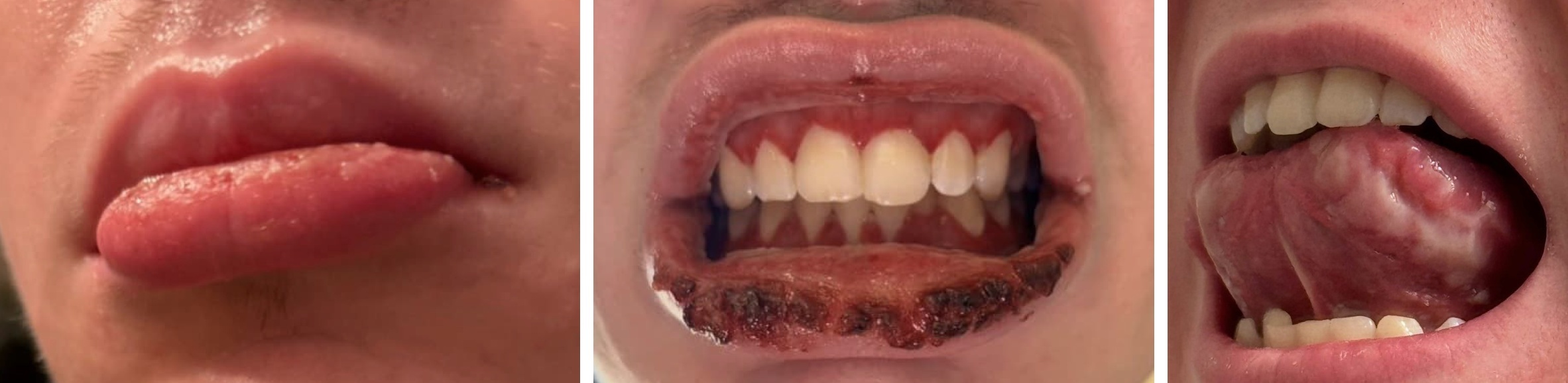
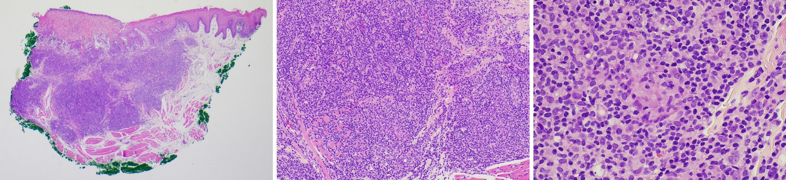
A previously healthy 20-year-old male presented with acute hypoxic respiratory failure following travel to a high-altitude area. Initial evaluation suggested multi-lobar pneumonia. The patient underwent bronchoscopy and an infectious workup, revealing positive urinary streptococcal antigen, positive Mycoplasma pneumoniae IgG antibodies, and positive herpes simplex virus type 1 and type 2 antibodies. Based on these findings, he was diagnosed with pneumococcal and mycoplasma pneumonia and treated with broad-spectrum antibiotics and high-dose systemic corticosteroids, resulting in significant clinical improvement. He was successfully extubated with complete resolution of his pulmonary symptoms; however, he continued to experience persistent fatigue. Several weeks later, the patient was re-hospitalized with oral mucositis consisting of palatal ulceration, swelling, desquamation, and ulceration of the lips (Figure 1). Following a subsequent recurrent episode of diffuse oral ulceration, the patient’s lateral and ventral tongue became involved and exhibited hyperplasia with a fissured appearance and areas of ulceration (Figure 1c). The lateral tongue was biopsied, which revealed non-necrotizing granulomatous inflammation (Figure 2), which was negative for infectious etiologies with GMS and AFB, thus pointing toward an underlying immune-mediated systemic granulomatous disease.
**_lip_edema._**(b)**_lip_ulceration_with_hemorrhagic_crusts_and_gingival_erythema._*.jpg)
The patient was treated with a steroid taper with complete resolution of his symptoms within two weeks. Notably, while receiving high-dose systemic corticosteroids for the oral ulcers, the patient reported no pulmonary symptoms. However, following the tapering of prednisone to 5 mg daily, the patient developed progressive dyspnea, headaches, and chest pain. Escalation of prednisone to 20 mg daily resulted in the prompt resolution of his symptoms.
**_a_wedge_of_formalin-fixed_mucosa_surfaced_by_parakeratotic_stratified_squamous_epi.jpeg)
During his hospitalizations, the patient’s pertinent laboratory workup is summarized in Table 1, with the most significant findings being hypogammaglobulinemia and cytopenia. Lymphocyte subset analysis demonstrated leukopenia with selective T-cell lymphopenia (reduced CD3+ and CD4+ cells) and preserved B-cell numbers, suggesting a predominantly T-cell–driven immunologic process.
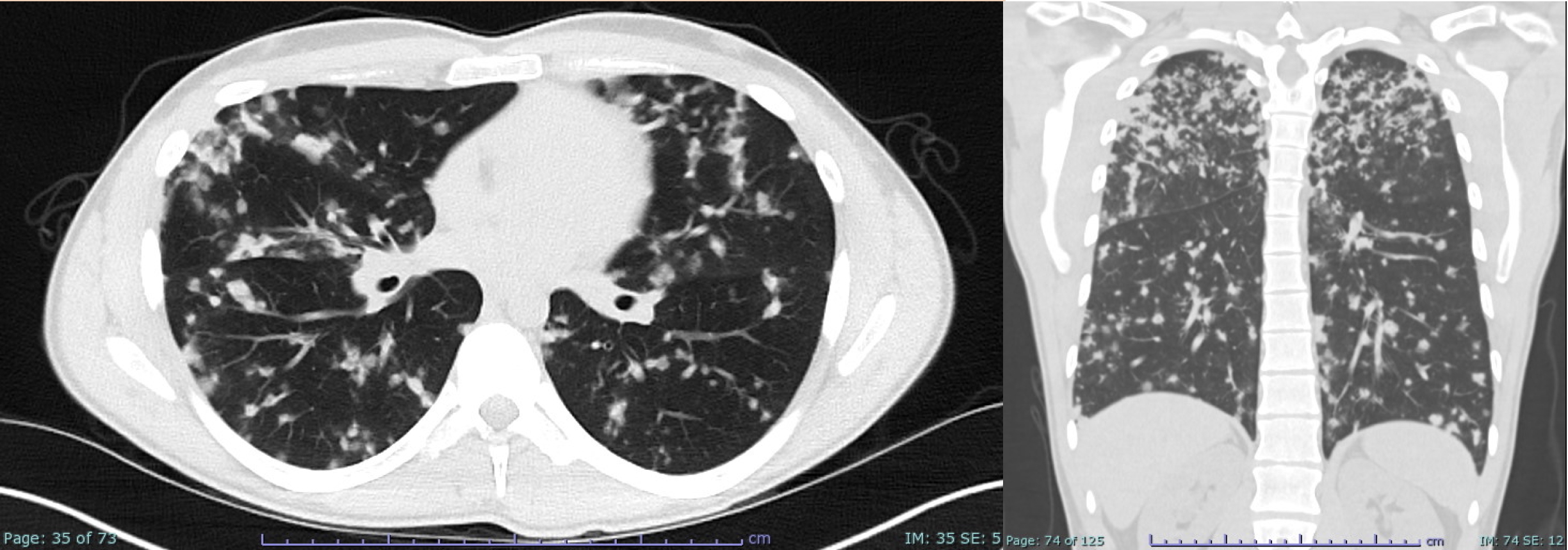
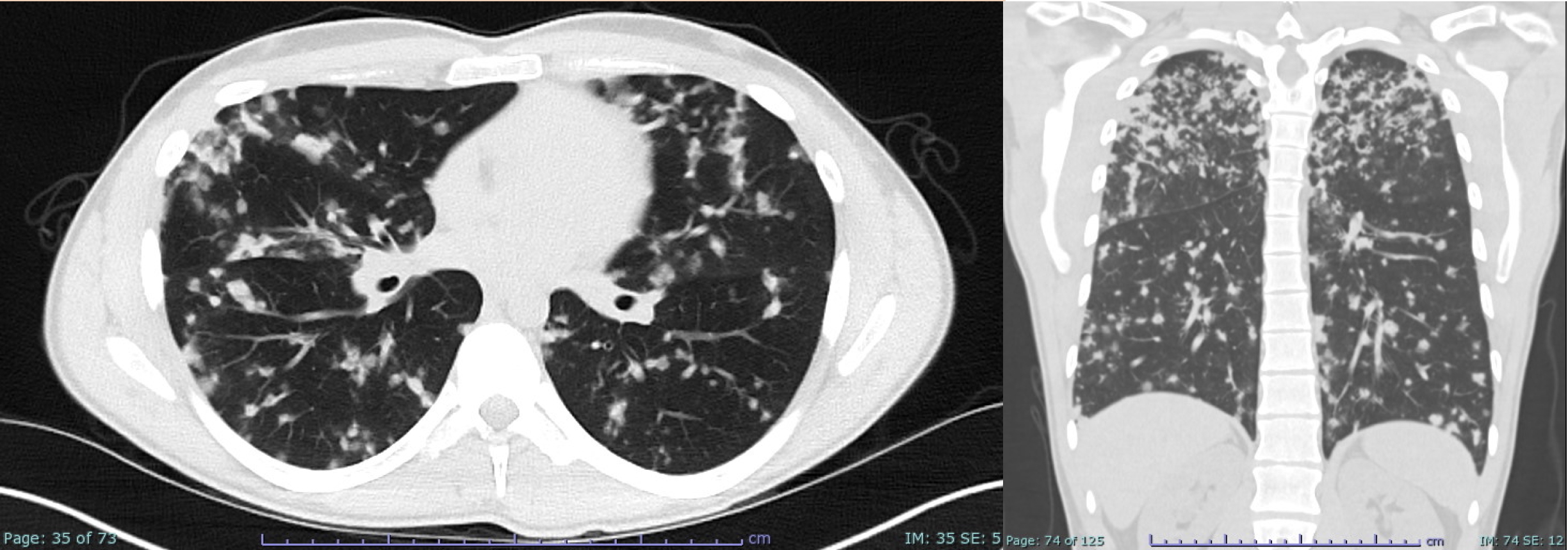
Serial chest imaging (Figure 3) demonstrated a dynamic, waxing and waning pattern of diffuse bilateral nodular airspace disease, initially improving with glucocorticoid therapy and recurring during steroid taper. The distribution evolved from predominantly upper lobe involvement to more diffuse perilymphatic nodularity affecting both upper and lower lobes. Transient right middle lobe consolidation with bronchial cutoff suggested superimposed airway-centered inflammation. Overall, the imaging strongly supported a steroid-responsive inflammatory granulomatous process, although features were not entirely classic for GLILD.
Additional findings included borderline splenomegaly and mild hepatomegaly with no lymphadenopathy. Pulmonary function testing showed near-normal lung volumes with mild airflow limitation and preserved diffusion capacity, without significant bronchodilator response.
Histopathologic examination of the lung biopsy demonstrated patchy, dense lymphoid infiltrates composed predominantly of small lymphocytes with relatively sparse plasma cells, centered around vascular structures and associated with foci of organizing pneumonia. B- and T-cell clonality studies supported a predominantly T-cell–driven process, and advanced immunohistochemical and molecular analyses showed no evidence of a lymphoproliferative disorder. Collectively, these findings were thought to be characteristic of immunodeficiency-associated lung disease and supported a diagnosis of granulomatous lymphocytic interstitial lung disease (GLILD) in the appropriate clinical context.
Immunologic evaluation did not establish a definitive diagnosis of common variable immunodeficiency (CVID), though hypogammaglobulinemia with pan-lymphopenia was identified. Vaccine titers were not contributory given confounding by prior intravenous immunoglobulin administration and concurrent glucocorticoid therapy. Genetic testing revealed a heterozygous NOD2 variant (p.Gly908Arg), which is not independently causative of primary immunodeficiency but may function as a disease modifier. Hematology deferred further evaluation given steroid-responsive cytopenias and the absence of significant organomegaly.
The patient was initiated on intravenous immunoglobulin (IVIg) therapy with a target serum IgG level of ≥700 mg/dL. Given the underlying immune dysregulation and steroid dependence, combination immunosuppressive therapy with rituximab and mycophenolate mofetil is planned to achieve targeted modulation of B- and T-cell activity as a steroid-sparing strategy. Age-appropriate vaccinations will be administered as indicated, and comprehensive infection-prevention measures will be implemented.
DISCUSSION
A systematic evaluation is essential to determine whether a patient has CVID or a CVID-related disorder. This includes a detailed personal and family history of immunodeficiency, recurrent sinopulmonary or gastrointestinal infections, and consideration of monogenic causes of CVID-like disease. Laboratory evaluation should confirm hypogammaglobulinemia, ideally outside of acute infections, and impaired responses to both protein and polysaccharide vaccines to confirm functional antibody deficiency.7 Secondary causes of hypogammaglobulinemia should be excluded, and additional studies, such as B-cell immunophenotyping to assess memory B-cell deficits, may be necessary.
Clinical, radiographic, and physiologic features of CVID-ILD often overlap with other ILDs, requiring diagnostic caution. Noninfectious otitis and sinusitis can occur in granulomatosis with polyangiitis, while CVID-associated enteropathy may cause malabsorption, chronic diarrhea, and nutritional deficiencies, resembling celiac disease or IBD. GLILD can mimic sarcoidosis, and splenomegaly or lymphadenopathy may precede lung involvement. Lymphoid hyperplasia in CVID can also mimic lymphoma radiologically. Autoimmune cytopenias are also common in other autoimmune disorders.8–10
On HRCT, CVID-ILD typically affects the mid to lower lungs, showing multiple small, smooth-margined nodules, patchy ground-glass opacities, and an airway-centric pattern. Bronchiectasis and airway disease are frequent and help distinguish CVID-ILD from other granulomatous lung diseases.11 In this case, lower lobe involvement, patchy ground-glass opacities, areas of consolidation, and smooth-margined nodules strongly favored GLILD or CVID-ILD. Histopathologic correlation further supports this distinction: patchy interstitial and perivascular lymphoid infiltrates, diffuse lymphoid hyperplasia, and organizing pneumonia favor CVID-ILD, whereas sarcoidosis is characterized by non-necrotizing epithelioid granulomas with a perilymphatic distribution and CD4⁺ T cell–predominance on BAL. These findings, while highly suggestive, require integration with clinical and immunologic data given overlap with other granulomatous lung diseases.
Not all cases of GLILD or CVID-ILD require treatment; therapy is generally reserved for symptomatic or progressive disease. IVIg significantly improves survival and quality of life by reducing infections, but approximately 70% of patients still develop noninfectious complications, most commonly chronic lung disease. IVIg should be optimized before starting immunosuppressive therapy to minimize respiratory infections and treatment-related complications.12 Systemic corticosteroids are first-line for remission induction in CVID-ILD/GLILD, while steroid-sparing agents—including mycophenolate, azathioprine, rituximab, abatacept, belimumab, or hematopoietic stem cell therapy—are reserved for select patient populations.13–15 Given the diagnostic complexity, overlap with other disease processes, and therapeutic implications, a multidisciplinary approach involving radiology, genetics, oncology, allergy–immunology, and hematology is recommended in the evaluation of these patients.
CONCLUSION
This case is significant because GLILD represented the initial manifestation of CVID. GLILD should be considered in young patients with steroid-responsive ILD, even in the absence of recurrent infections, and that extrapulmonary granulomas alone are insufficient to confirm sarcoidosis. The presence of autoimmune cytopenias, hypogammaglobulinemia, and T- cell lymphopenia indicated immune dysregulation consistent with GLILD. Multidisciplinary care with pulmonology, immunology, rheumatology, radiology, and clinical pathology is often needed to secure an accurate diagnosis.