
Introduction
A miliary pulmonary pattern, characterized by innumerable 1–3 mm micronodules distributed throughout the lungs represents one of the most distinctive radiographic findings encountered in pulmonary medicine. Although clinicians most often associate this pattern with disseminated infections such as miliary tuberculosis, systemic fungal infections (including histoplasmosis, coccidioidomycosis, cryptococcosis, and blastomycosis), nocardiosis, viral infections in immunocompromised hosts and septic emboli, non-infectious etiologies represent a substantial proportion of cases. These include granulomatous diseases like sarcoidosis and hypersensitivity pneumonitis, occupational lung diseases such as silicosis and coal workers’ pneumoconiosis, and metastatic malignancies.
Among malignant causes, MIPC constitutes a rare yet clinically meaningful presentation of lung adenocarcinoma, accounting for approximately 1–2% of non–small cell lung cancer (NSCLC) cases.1 Its diffuse micronodular pattern closely mimics infection, contributing to diagnostic delay, inappropriate antimicrobial therapy, and postponed oncologic evaluation. MIPC is strongly associated with EGFR mutations, especially in younger never-smokers, making early biopsy and molecular testing essential. As Choi et al implied, the frequency of EGFR mutations can be as high as 58% in MIPC compared to 24% in non-MIPC NSCLC.2 Prompt initiation of EGFR-targeted TKIs can significantly improve outcomes compared with conventional chemotherapy.
Case Report
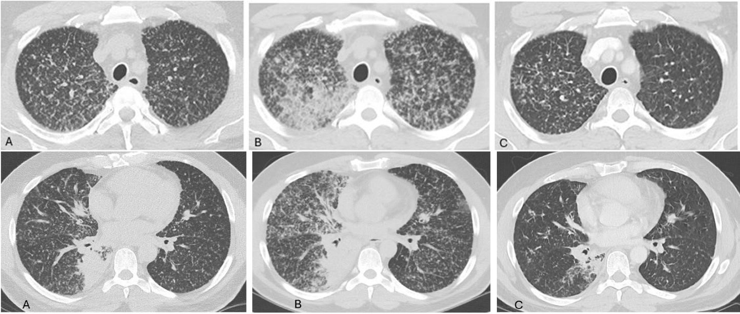
A 43-year-old Caucasian male never-smoker living in Arizona presented with three weeks of worsening dyspnea and a non-productive cough. Initial chest imaging showed diffuse bilateral
miliary nodularity (Figure 1A). Despite three negative acid-fast bacilli smears, negative coccidioidomycosis screening, and the absence of systemic symptoms, he was treated empirically for presumed infectious causes.
**_initial_presentation._computed_tomography_of_upper_(top_panel)_and_lower_(bottom_p.jpeg)
Over the next four weeks, his respiratory status deteriorated significantly. Pulmonary function testing demonstrated a new restrictive ventilatory and gas exchange defect. Repeat chest computed tomography revealed progression of the miliary pattern (Figure 1B), prompting transbronchial and endobronchial ultrasound–guided biopsies. Histopathology of right lower lobe lung tissue and station 4 left lower paratracheal lymph node confirmed moderately differentiated lung adenocarcinoma, and molecular analysis identified an EGFR mutation. Positron emission tomography showed no evidence of extrapulmonary metastasis at diagnosis.
He was started on EGFR-targeted therapy with osimertinib alongside pemetrexed and carboplatin. Five months after initiating EGFR-targeted treatment, he exhibited marked clinical and radiographic improvement (Figure 1C). He was able to resume normal daily activities and return to hobbies, including mountain hiking, that he had been unable to perform prior to therapy.
Discussion
This case highlights the risk of diagnostic anchoring when a miliary nodular pattern is assumed to be infectious despite the absence of classic risk factors. The delayed recognition of MIPC postponed appropriate oncologic evaluation. Fortunately, this patient’s tumor harbored an EGFR mutation, allowing effective treatment with a targeted TKI and resulting in substantial clinical and radiographic improvement.
A miliary nodular pattern reflects hematogenous dissemination rather than a single specific diagnosis, with diffuse micronodular spread throughout the lungs.3 Clinicians must avoid prematurely attributing this pattern solely to infectious causes and instead maintain a broad differential that includes non-infectious conditions. As shown by Salahuddin et al., approximately half of miliary presentations are non-infectious, including sarcoidosis (24%), silicosis (14%), extra-thoracic malignancies metastatic to the lung (10%), and primary lung cancer (1%).4 Although malignancy represents roughly 10% of miliary patterns, failure to consider it early can lead to inappropriate antimicrobial therapy, delays in cancer diagnosis, and increased mortality. MIPC can be highly aggressive and accounts for approximately 1-2% of NSCLC.1 Untreated metastatic NSCLC carries a median survival of only approximately 7 months.5 Anchoring on infection can therefore squander a critical window in which early oncologic intervention may improve survival.
A distinguishing feature of MIPC is its strong association with EGFR mutations: 70–91% of patients with this radiographic phenotype harbor an EGFR alteration, far exceeding the 15–50% prevalence in lung adenocarcinoma overall.6 Early biopsy and molecular testing are therefore essential, as timely identification and treatment can prevent further metastatic spread and significantly improve outcomes. EGFR-targeted TKIs markedly extend survival in EGFR-mutated MIPC, with median overall survival of 17.8 months compared versus 10.6 months in patients without the EGFR mutations. When initiated early and in the absence of extrapulmonary metastasis, survival can increase to 23.5 months versus 10.4 months in those with metastatic disease.7 These data underscore EGFR mutation status as a key prognostic marker and EGFR inhibitors as highly effective therapy for this cancer phenotype.
Although EGFR mutations occur more frequently in certain populations (approximately 50–60% of Asians and 15–20% of Caucasians with lung adenocarcinoma),7 there are no statistically significant racial differences in overall survival among those treated with EGFR-targeted therapy, especially when a newer generation TKI is used.8 Lower survival reported in black patients with EGFR-mutated NSCLC appears multi-factorial with confounding factors such as higher rates of decision to pursue hospice and inability to attend oncology visits due to frequent hospitalizations.9 To this point, a more recent retrospective study in 2024 showed no statistically significant difference in overall survival among Asian, Black, and White populations, especially when first-line TKI therapy with osimertinib was used.8 Because the therapeutic benefit is highly effective across all racial groups, recognizing a miliary pattern should prompt early oncologic referral and timely initiation of targeted therapy to maximize benefit for all patients.
Conclusion
A miliary pulmonary pattern often reflects hematogenous dissemination of disease and requires a broad differential diagnosis that includes infectious and noninfectious etiologies. Clinicians should avoid diagnostic anchoring and pursue timely tissue diagnosis when the clinical picture is atypical. In never-smokers, primary lung adenocarcinoma presenting as MIPC should be considered, as early molecular testing and initiation of EGFR-targeted therapy can significantly improve outcomes and survival.