
Introduction
Granulomatous-lymphocytic interstitial lung disease (GLILD) is a form of interstitial lung disease characterized histopathologically by non-caseating granulomas surrounded by lymphocytic interstitial infiltrates or surrounding a reactive follicle.1 While GLILD is most often encountered in patients with underlying CVID, rare cases have been reported in patients without the classic CVID profile.2 Early identification using computed tomography (CT) imaging in conjunction with multidisciplinary evaluation and targeted biopsy allows for differentiation of GLILD from more prevalent mimics such as sarcoidosis, hypersensitivity pneumonitis, lymphoid interstitial pneumonia,3,4 granulomatous infections,5 and pulmonary lymphoproliferative disorders.6
Case
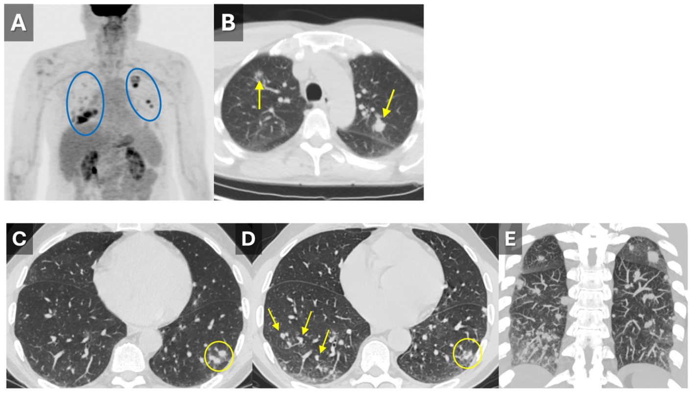
A 65-year-old man was referred to our pulmonary service by ENT for further evaluation of multiple pulmonary nodules. The patient had initially presented to ENT with left nasal congestion. Imaging with sinus CT and brain MRI revealed a left nasal cavity mass and a PET-CT was ordered. Although the nasal mass was eventually diagnosed as a benign inflammatory polyp, the PET-CT revealed multiple incidental FDG-avid pulmonary nodules (Figure 1A,B). The patient’s past medical history included hypothyroidism, hyperlipidemia, and dysphagia. He had a 10-year history of smoking a few cigarettes per day but quit 40 years ago and had no occupational or environmental exposures. He denied dyspnea or constitutional symptoms at presentation and did not have a history of symptoms to suggest any recurrent infections or connective tissue disease.
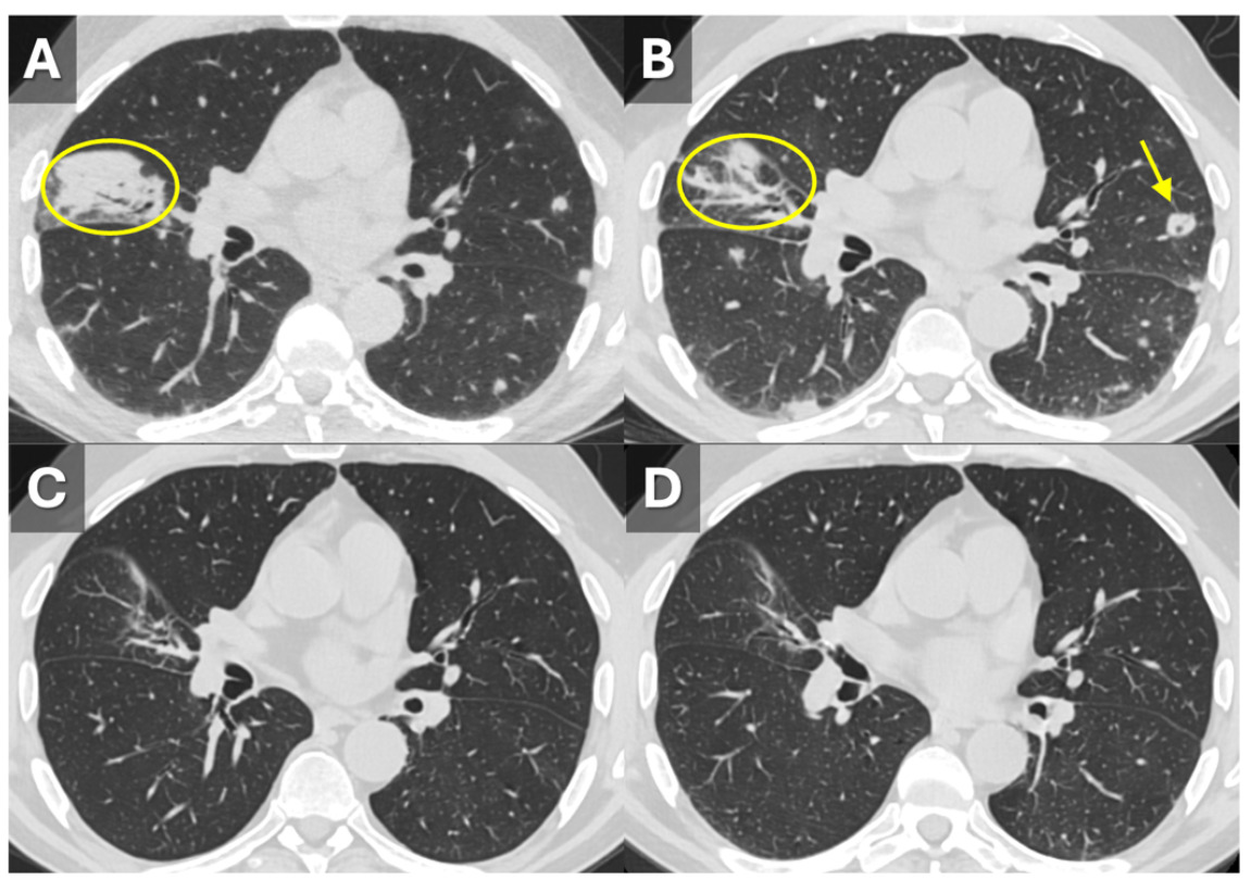
A diagnostic CT of the chest was performed with subsequent CT-guided biopsy, the results of which showed organizing acute lung injury in a background of non-necrotizing granulomatous inflammation. Infectious disease was consulted, and broad-spectrum antimicrobial therapy, including antifungal coverage with Posaconazole, was initiated. However, extensive infectious disease workup was negative. A follow-up CT performed ~1 month later showed improvement in some of the initial CT findings; however, several new nodules of various size appeared, some of which were partially cavitary. Findings were consistent with spontaneous waxing and waning pulmonary findings rather than progressive infection or malignancy (Figure 1C-E, Figure 2A-B). The nodules generally appeared to favor a peribronchial/perivascular distribution, although some were peripheral, located along the pleura.
A wedge biopsy of the right upper lobe was performed and again showed organizing acute lung injury in a background of extensive non-necrotizing granulomatous inflammation. Immunohistochemical stains for CD2, CD3, CD4, CD5, CD7, CD8, CD10, CD15, CD20, CD21, CD30, CD43, BCL2, BCL6, and cyclin D1 demonstrated a mixed population of predominantly small T and B cells with preserved CD4/CD8 expression and no aberrant expression profile, ruling out lymphoproliferative disease. Acid-fast (AFB) and fungal (GMS) stains were negative and a diagnosis of GLILD was made.
Comprehensive immunologic evaluation showed mild lymphopenia, with a CD4 count of 460 cells/ul, CD8 count of 65 cells/ul, and normal CD4:CD8 ratio. Immunoglobulin levels revealed normal IgG, IgA, IgM, and IgG4 subclasses. Genetic testing for CTLA-4 mutations was negative.
The patient was started on prednisone 1 mg/kg body weight (40 mg daily) for GLILD. The patient was started on trimethoprim-sulfamethoxazole prophylaxis while on immunosuppression with a slow steroid taper over approximately 14 weeks, with consideration of rituximab and mycophenolate mofetil in the future.
Multidisciplinary evaluation involving pulmonary, radiology, immunology, pathology, infectious disease, and hematology specialists was essential for establishing the correct diagnosis. Pulmonary function testing at initial evaluation showed mildly reduced diffusing capacity. Repeat pulmonary function testing several months after initiation of corticosteroid therapy demonstrated slightly improved diffusing capacity. Serial imaging demonstrated near-complete resolution of pulmonary findings (Figure 2C-D), concordant with the patient’s clinical improvement.
_coronal_fdg-pet_ct_demonstrating_many_fdg-avid_pulmonary_nodules_(circled)__and_maximu.png)
__and_follow-up_ct_1_month_later_(b)__and_at_3_mon.png)
Discussion
GLILD represents a rare non-infectious granulomatous-lymphoproliferative lung disorder, which is usually seen in association with CVID, but can rarely manifest outside the clinical context of CVID.1 GLILD complicates approximately 8–20% of CVID cases,3 whereas its prevalence outside of a diagnosis of CVID is unclear due to the rarity of cases reported, but has been described in genetic conditions such as 22q11.2 deletion syndrome (DiGeorge syndrome)2 as well as deficiency of lipopolysaccharide (LPS) responsive beige-like anchor protein (LRBA), cytotoxic T-lymphocyte antigen-4 (CTLA4), Kabuki syndrome, and hyperactive signal transducer and activator of transcription 3 (STAT3).7
The pathogenesis of GLILD is not entirely understood but speculated to be due to autoreactive or chronically stimulated T cells with a subsequent dysregulated cytokine response, leading to unopposed lymphoid proliferation in pulmonary tissues.2 This lymphoproliferation may be further dysregulated in GLILD due to an absence of FoxP3+ regulatory T cells.2 Abnormal response to human herpes virus-8 (HHV-8) has also been hypothesized to play a role.2,8
Symptoms are variable and often insidious and non-specific, most commonly including exertional dyspnea, nonproductive cough, fatigue, and other constitutional symptoms.1,2,4,5 Patients may be asymptomatic, including during radiographic relapse.2 Systemic signs such as splenomegaly and lymphadenopathy may be present, particularly in those with immunodeficiency.5
CT most commonly reveals multiple, small (<10 mm) nodules that are centrilobular or randomly distributed, low-grade bronchiectasis associated with fibrosis, reticulation, and ground-glass opacities that are often predominantly in the lower lobes.1,2,5 This differs from sarcoid, which predominantly affects the middle-upper lung fields, and less commonly involves bronchiectasis, which is mainly traction bronchiectasis when present.1 Enlarged hilar and mediastinal lymph nodes are frequently observed in GLILD, which overlaps with sarcoidosis.1,5
Diagnosis can be difficult due to the variable, nonspecific presentation. Multidisciplinary evaluation is recommended, and referral to an interstitial lung disease (ILD) center is preferred in cases of diagnostic uncertainty.4 Treatment currently lacks established, evidence-based guidelines.6 Initial management typically involves corticosteroids to suppress granulomatous and lymphoid inflammation.9 Immunomodulatory therapy such as azathioprine and mycophenolate mofetil, as well as rituximab, may be used as corticosteroid-sparing agents in refractory or relapsing cases.4,9
Recognition of GLILD in the absence of CVID is critical to avoid misdiagnosis and potential unnecessary treatments for presumed infection, malignancy, or systemic vasculitis.2–4,8,9 In this case, the waxing and waning pattern of nodules on serial imaging, combined with the clinical and radiologic improvement with corticosteroid therapy, was inconsistent with progressive infection or malignancy and strongly supported an immune-mediated process. In some cases, genetic testing for mutations associated with primary immunodeficiencies (such as CTLA-4, LRBA, STAT3) may eventually identify a specific genetic basis for the immune dysregulation, potentially enabling targeted therapy. Suspicion of GLILD by clinicians and radiologists will allow for timely diagnosis and appropriate treatment.