
Introduction
Bilateral pneumothoraces after double-lung transplantation is an uncommon but recognized complication. In most individuals, the right and left pleural cavities are separate. However, in many double-lung transplant recipients, the functional anatomy of the mediastinum has been altered postoperatively, resulting in a single, interpleural communication. A communicating space and resulting complications, such as communicating pneumothorax, have been commonly described in the immediate post-operative period. However, persistence years after transplant has been less well observed and characterized. The presence of a communicating pleural space has significant clinical implications because air or fluid introduced into one hemithorax will extend to the opposite side, potentially resulting in bilateral pneumothorax and placing patients at high risk for severe hypoxemia and hemodynamic instability.1,2 This case report details the presentation and management of bilateral pneumothoraces in a double-lung transplant recipient.
Case Report
An 80-year-old man with a past medical history of idiopathic pulmonary fibrosis status post bilateral lung transplant via clamshell incision in 2020, chronic hypoxic respiratory failure requiring supplemental oxygen at 3 liters per minute via nasal cannula at baseline, recurrent deep vein thrombosis on apixaban, hypertension, hyperlipidemia, cutaneous pleomorphic dermal sarcoma of the scalp, and heart failure with preserved ejection fraction presented to the emergency department with one week of persistent right scapular pain following a mechanical fall from a chair onto his right side. He denied head trauma, additional injuries, or dyspnea at the time of the fall. His only new symptom was progressive right scapular pain in the days afterward. He reported chronic shortness of breath consistent with baseline and noted several episodes of aggressive coughing over the preceding two weeks.
Prior to this presentation, the patient had been followed for chronic lung allograft dysfunction (CLAD). Spirometric evaluation demonstrated progressive airflow obstruction over several years, with the post-transplant FEV1 baseline (the mean of the two highest post-transplant values) calculated to be approximately 3.27 L. At the time of presentation, the most recent spirometry (September 2025) demonstrated an FEV1 of 0.75, which was 23% of the post-transplant baseline, consistent with definite CLAD Stage 4 per the 2019 International Society for Heart and Lung Transplantation (ISHLT) consensus criteria.3 Spirometry demonstrated mixed obstructive and restrictive physiology (FEV1/FVC 58%, FVC 41% of predicted). Transbronchial biopsies performed in December 2022 and April 2025 showed no acute or chronic rejection (ISHLT Grade A0/B0/C0 and A0/BX, respectively), and CT-based CLAD phenotyping was not conclusively established. The patient had completed salvage radiation therapy (60 Gy in 20 fractions) for recurrent multifocal scalp pleomorphic dermal sarcoma three weeks prior to this presentation.
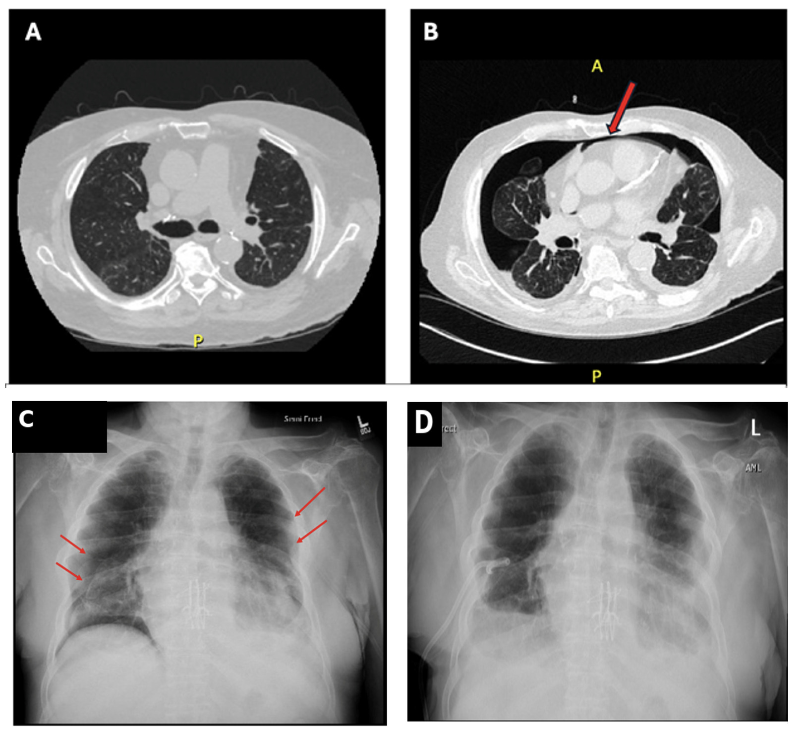
On presentation, the patient was hemodynamically stable with appropriate oxygen saturation on 3 L of oxygen via nasal cannula. Laboratory studies were unremarkable. Computed tomography (CT) of the chest revealed bilateral pneumothoraces (right greater than left), trace-to-small bilateral pleural effusions, and a new pulmonary nodule (Figure 1A). Given the presence of a communicating pleural space on imaging, the pulmonary team elected to place a right-sided chest tube to facilitate re-expansion of both lungs. Apixaban was held for 24 hours, and the patient was transitioned to intravenous heparin for anticoagulation, which was discontinued four hours prior to the procedure.
**_ct_scan_from_8_months_prior_to_admission_showing_no_evidence_of_pneumothorax._**(b.png)
A right-sided 14-French pigtail catheter chest tube was inserted percutaneously using the Seldinger technique (Wayne Pneumothorax Set) under real-time ultrasound guidance. The pneumothoraces appeared free-flowing on CT without evidence of loculation, permitting single-tube decompression. The lung transplant team was notified of the hospitalization and treatment plan; no additional recommendations were provided. The chest tube was placed on suction. A follow-up chest radiograph demonstrated resolution of both the right and left pneumothoraces. After 24 hours of suction, the chest tube was placed to water seal for three hours and then clamped. Serial chest radiographs demonstrated successful bilateral lung re-expansion (Figure 1B). On hospital day 3, the chest tube was removed, and the patient was observed for 6 hours prior to discharge. At the time of discharge, he reported improved shortness of breath and improved right scapular pain. Following discharge, repeat chest CT demonstrated interval progression of the pulmonary nodule (Figure 1C), prompting robotic bronchoscopy. Histopathologic analysis confirmed metastatic pleomorphic dermal sarcoma, likely originating from his known scalp lesion.
Discussion
This case report describes a patient who developed bilateral pneumothoraces secondary to a shared pleural cavity—an anatomical alteration known as “buffalo chest,” commonly observed after double-lung transplantation via clamshell incision.4 Recognition of a communicating pleural space is critical, as air introduced into one hemithorax can traverse to the opposite side, resulting in bilateral pneumothoraces and placing the patient at risk for rapid hypoxemia and hemodynamic instability. Interestingly, despite the presence of bilateral pneumothoraces, this patient remained hemodynamically stable and did not exhibit hypoxia or respiratory distress, suggesting a more chronic process.
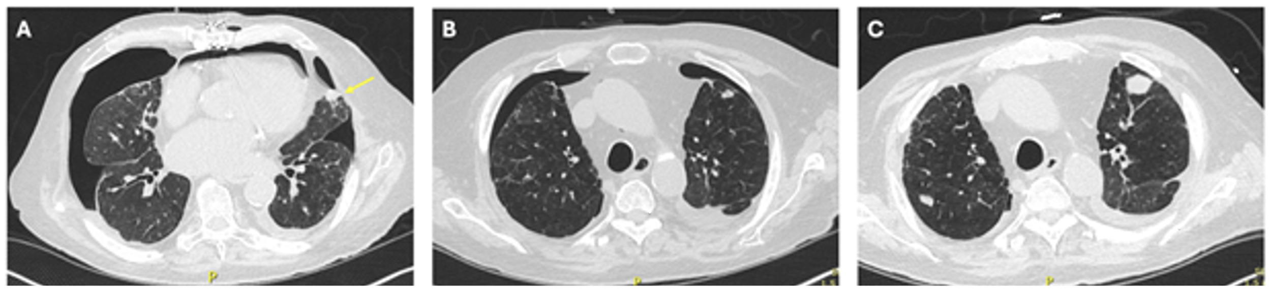
The etiology of the pneumothoraces in this patient is unknown, but likely multifactorial. The presenting mechanical fall onto his right side may have caused pleural or parenchymal injury, leading to an air leak. Episodes of forceful coughing over the preceding two weeks may have further increased intrathoracic pressure, predisposing him to rupture of subpleural blebs—a mechanism of particular relevance given his advanced CLAD. Patients with obstructive CLAD (bronchiolitis obliterans syndrome) demonstrate significant air trapping and thoracic hyperinflation analogous to advanced emphysema, creating peripheral hyperlucent lung zones at increased risk for air leak following trauma or acute airway pressure elevations.5 Metastatic sarcoma may also likely have contributed. Peripheral pulmonary metastases can precipitate spontaneous pneumothorax through several mechanisms, including direct visceral pleural invasion, tumor necrosis with bronchopleural fistula formation, and intermittent bronchiolar obstruction by subpleural nodules leading to distal bleb formation.6,7 In this patient, a progressing subpleural sarcoma deposit (Figure 2) represents a plausible contributor via direct pleural disruption, potentially exacerbated by radiation-induced tumor necrosis following the recently completed salvage radiotherapy.
**_initial_axial_ct_on_presentation_demonstrating_a_metastatic_lesion_(arrow)_with_bi.png)
The finding of a communicating pleural space justified the decision to place a single right-sided chest tube, with the expectation that decompression would allow for bilateral lung re-expansion. This strategy also reduced procedural risk, including infection and bleeding, and improved patient comfort. The pneumothoraces appeared free-flowing on imaging without loculation, which facilitated this approach. It should be noted, however, that pleural adhesions from post-surgical scarring are common in patients years after transplantation and may result in trapped or loculated pneumothoraces that require alternative management—underscoring the importance of pre-procedural ultrasound assessment in this population. This case contributes to the limited body of literature describing successful resolution of bilateral pneumothoraces using a single chest tube in patients with a post-transplant communicating pleural space.8,9 However, this strategy remains relatively uncommon and is not specifically addressed in current clinical guidelines, highlighting the need for further research to better define its safety and efficacy.
Finally, this case illustrates that new or unexplained pneumothorax in a double-lung transplant recipient, particularly in the absence of clear traumatic or physiologic explanation, may prompt careful evaluation for occult malignancy. Post-transplant malignancy, including pleomorphic dermal sarcoma and other mesenchymal tumors, is a well-recognized complication of chronic immunosuppression, and pulmonary metastases from these malignancies are an underappreciated cause of spontaneous pneumothorax in this population.6,7