
Introduction
Progressive pulmonary fibrosis (PPF) is a diagnostic entity describing interstitial lung disease (ILD) with a progressive phenotype independent of the underlying etiology. The 2022 definition proposed by Raghu et al. requires at least two of three criteria to occur within one year in a patient with ILD other than idiopathic pulmonary fibrosis (IPF), without an alternative explanation and despite appropriate management: (1) worsening respiratory symptoms (e.g., increasing dyspnea or cough); (2) physiologic evidence of disease progression, defined as an absolute decline in forced vital capacity (FVC) of ≥5% predicted or an absolute decline in diffusing capacity for carbon monoxide (DLCO) of ≥10% predicted within one year; and (3) radiologic evidence of progression on high-resolution CT imaging, including increased fibrosis, worsening traction bronchiectasis, or increased reticulation or honeycombing.1 Progressive fibrotic lung disease may complicate a variety of non-IPF ILDs, including connective tissue disease–associated ILD, hypersensitivity pneumonitis, and granulomatous lung diseases.1 Identification of the underlying etiology is essential, as management may involve immunomodulatory therapy, antifibrotic agents, or a combination of both.1
Hypereosinophillic syndrome (HES) is a heterogeneous group of disorders characterized by persistent eosinophilia and eosinophil-mediated end-organ damage. Pulmonary involvement is common, typically manifesting as eosinophilic pneumonia or thromboembolic disease; however, progression to PPF-ILD is rare. Management depends on disease subtype, with corticosteroids as first-line therapy and refractory cases requiring agents such as imatinib, azathioprine, or the interleukin-5 antagonist mepolizumab.2 We present a case of severe HES complicated by progressive pulmonary fibrosis and pulmonary hypertension, ultimately requiring combined heart–lung transplantation.
Case Report
A 44-year-old man with a long-standing history of hypereosinophilic syndrome presented with progressive dyspnea. His medical history was notable for an eosinophilic intracardiac mass complicated by endocarditis and severe aortic insufficiency requiring mechanical aortic valve replacement 20 years earlier, at which time bone marrow biopsy confirmed HES. Initial therapy with imatinib (dose unknown) resulted in no clinical response. He subsequently required intermittent corticosteroids and long-term azathioprine for disease control. Due to ongoing disease activity, mepolizumab 300 mg monthly was initiated but discontinued after worsening dyspnea.
Several months prior to presentation, the patient was hospitalized for acute hypoxic respiratory failure with a white blood cell count of 7.8 × 10³/µL and eosinophil count of 300/µL. Chest radiography demonstrated diffuse bilateral interstitial infiltrates. He was treated with prednisone 40 mg daily, followed by a gradual taper, and discharged on 3 L/min of supplemental oxygen.
He later presented to the interstitial lung disease clinic with worsening dyspnea and increased oxygen requirements (6 L/min). Physical examination revealed bibasilar coarse crackles, and oxygen saturation was 90% on 4 L/min via nasal cannula. Laboratory evaluation showed a white blood cell count of 8.9 × 10³/µL with eosinophils of 480/µL and elevated lactate dehydrogenase (1,099 U/L). Notable social history includes a lifelong nonsmoking status, prior military service, and no known toxic or burn pit exposures.
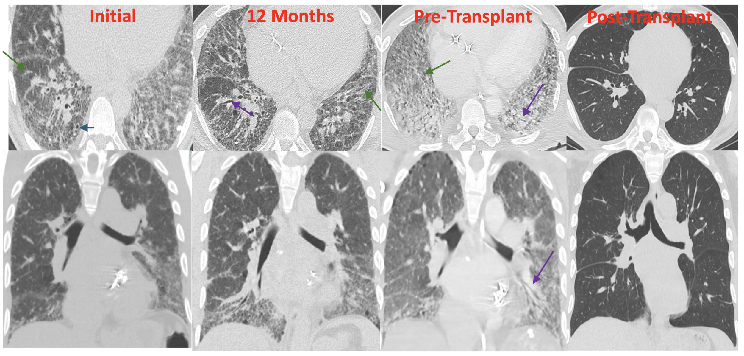
High-resolution computed tomography of the chest demonstrated fibrotic interstitial lung disease with subpleural reticulation, architectural distortion, and traction bronchiectasis in a lower lobe predominance with significant ground glass, indeterminate for UIP (Figure 1). Transthoracic echocardiography revealed preserved left ventricular systolic function (ejection fraction 60%) with right ventricular enlargement, reduced systolic function, and an estimated right ventricular systolic pressure of 88 mmHg. Comprehensive autoimmune serologies were negative, and serum IgE was elevated at 363 IU/mL. Pulmonary function testing demonstrated severe restriction with an FVC of 1.93 L (34% predicted), FEV₁ of 1.54 L (35% predicted), total lung capacity of 3.13 L (41% predicted), and preserved FEV₁/FVC ratio. DLCO could not be obtained due to hypoxemia. Six-minute walk distance was markedly reduced to 120 m, terminated early due to dyspnea.
_and_coronal_(bottom)_computed_tomography_(ct)_images_demonstra.png)
Following a multidisciplinary discussion involving radiology, cardiology, hematology, and pulmonology, the radiologic pattern was favored as representing NSIP, and the possibility of tissue biopsy was explored to help guide diagnosis and management. The patient was deemed too high-risk for a surgical lung biopsy. Bronchoscopy with bronchoalveolar lavage (BAL) was performed, demonstrating 61% neutrophils and 27% eosinophils. Cytology revealed scattered multinucleated giant cells without evidence of infection or malignancy. Bacterial, fungal, mycobacterial, and viral studies were negative. Endobronchial biopsies demonstrated squamous metaplasia without active inflammation.
High-dose prednisone (60 mg/day) was restarted but resulted in significant adverse effects, including edema, emotional lability, and steroid-induced diabetes. Repeat bone marrow biopsy showed no evidence of hypereosinophilia, although the patient was receiving prednisone at the time. Azathioprine was transitioned to mycophenolate mofetil but discontinued due to gastrointestinal intolerance, and azathioprine was resumed. Despite immunosuppression, the patient experienced progressive symptoms and radiographic worsening consistent with PPF-ILD. Pirfenidone was initiated and titrated to 801 mg three times daily but discontinued due to gastrointestinal intolerance, prompting referral for transplant evaluation.
Repeat cardiac evaluation demonstrated worsening pulmonary hypertension with right ventricular dysfunction. Right heart catheterization revealed a mean pulmonary artery pressure of 52 mmHg, pulmonary capillary wedge pressure of 25 mmHg, and pulmonary vascular resistance of 4.7 Wood units, consistent with combined pre- and post-capillary pulmonary hypertension. The patient rapidly deteriorated with refractory hypoxic respiratory failure, cardiac arrest, and cardiogenic shock due to biventricular failure, necessitating emergent transfer to a transplant center, and he successfully received combined heart–lung transplantation.
Discussion
Hypereosinophilic syndrome is defined by persistent eosinophilia (absolute eosinophil count ≥1,500 cells/µL) or eosinophilic tissue infiltration with associated organ damage, after exclusion of secondary causes such as infection, allergic disease, and malignancy.1 Evaluation includes bone marrow biopsy, molecular testing, and immunophenotyping to identify clonal or lymphocytic variants, with common mutations involving FIP1L1–PDGFRA, PDGFRB, FGFR1, and JAK2.1,3
Pulmonary involvement occurs in up to 70% of patients with HES and most commonly presents as eosinophilic pneumonia, ground-glass opacities, or pulmonary nodules.1,3 These manifestations typically respond to corticosteroids or targeted therapy. Treatment is guided by disease subtype, with imatinib preferred for PDGFRA/B-associated disease and mepolizumab demonstrating efficacy in idiopathic and lymphocytic variants.2,4 Progression to progressive pulmonary fibrosis–ILD, however, appears to be rare.1 Reported cases most often demonstrate non-specific interstitial pneumonia patterns that improve with immunosuppression.2 In contrast, our patient developed progressive fibrosis despite resolution of peripheral eosinophilia and aggressive therapy.
Eosinophil-driven fibrosis is mediated through multiple overlapping mechanisms. Eosinophils and Th2 lymphocytes secrete profibrotic cytokines, including transforming growth factor–β, interleukin-4, and interleukin-13, promoting fibroblast activation and extracellular matrix deposition.5 Eosinophil granule proteins cause direct epithelial injury, perpetuating aberrant wound repair, while chronic inflammation induces endothelial dysfunction, microvascular remodeling, and hypoxia-driven fibroblast proliferation.6 These processes may sustain fibrosis even in the absence of peripheral eosinophilia, as observed in this case.
To our knowledge, there are no published cases describing HES as a primary indication for lung or heart–lung transplantation. Post-transplant studies, however, demonstrate that eosinophilia is associated with increased rejection and chronic lung allograft dysfunction.7 This case expands the spectrum of HES-associated pulmonary disease, highlighting eosinophilic inflammation as a potential driver of progressive fibrotic lung disease and pulmonary hypertension refractory to standard therapy. Early recognition of fibrotic transformation and timely referral for advanced therapies may be critical in selected patients.