
Introduction
Innominate artery compression syndrome is a rare cause of airway obstruction that is commonly diagnosed in the pediatric population. This condition arises from a congenital vascular malformation. Given that this condition is seldom diagnosed in adulthood, this diagnosis can be missed.
Case Report
A 52-year-old female with a history of hypothyroidism, rheumatoid arthritis, bipolar disorder, morbid obesity, fibromyalgia, deep vein thrombosis, and asthma presented with progressive worsening of her chronic cough and new onset dyspnea. A recent admission included a laryngoscopy that noted vocal cord dysfunction. In the emergency department, the patient demonstrated stridor and rhonchi on physical exam. She received intravenous steroids and albuterol-ipratropium nebulizer treatments. Notably, the outpatient pulmonary function test two months prior to presentation showed no airflow limitation or significant bronchodilator response. There was symmetric reduction in FEV1 and FVC which may be related to body habitus or restrictive lung disease; however, no fixed airway obstruction or variable intrathoracic obstruction were noted on the flow-volume loop.
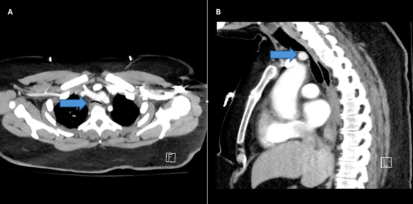
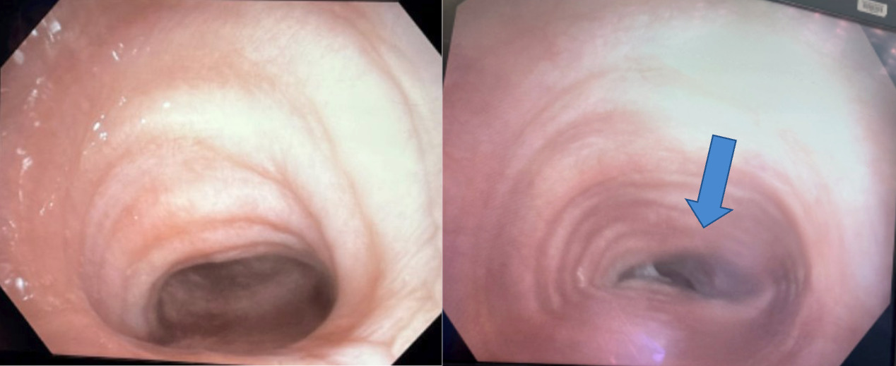
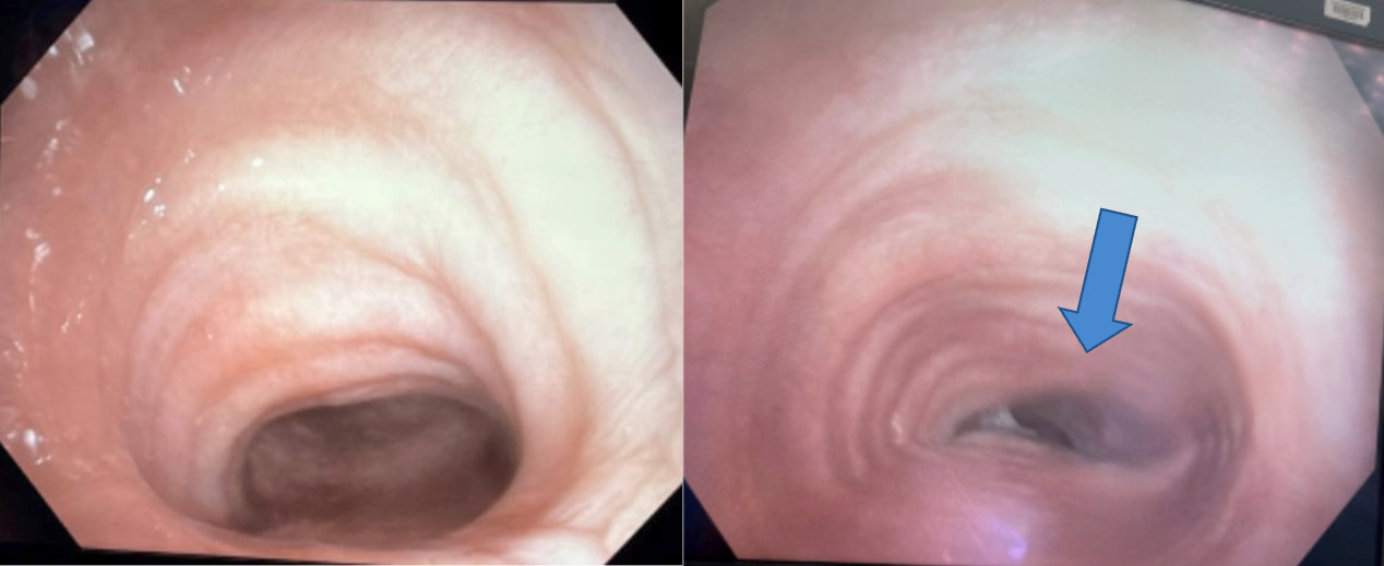
Computed tomography (CT) angiography of the chest was performed to evaluate for pulmonary embolism and revealed tracheal narrowing 4 cm superior to carina, raising concern for external compression by the innominate artery (Figure 1). Bronchoscopy confirmed significant pulsatile tracheal compression of the 5th-6th tracheal rings consistent with extrinsic vascular compression (Figure 2). Due to the high risk of vascular erosion, stent placement was deferred. The patient underwent surgical debranching of the innominate artery where the artery was divided at its origin from the aorta. The distal innominate artery was then connected to a graft and reimplanted to ascending aorta. Post-operative course was complicated by pericardial effusion causing tamponade requiring surgical pericardial window. The patient had a complicated post-operative course with persistent respiratory failure requiring prolonged intubation, likely in part due to persistent tracheomalacia of the trachea despite alleviation of compression. The patient underwent tracheotomy prior to being discharged to a rehabilitation facility and was able to safely decannulate a few months later.
_and_sagittal_(b)_reconstructions_from_a_contrast-enhanced_chest_ct_angiogram._an.png)
Discussion
Innominate artery compression syndrome occurs from an abnormally distal and posterior origin of the artery from the aortic arch.1 The artery crosses toward the right upper extremity and can compress the trachea as it ascends. In the symptomatic pediatric population, stridor, respiratory distress, or failure to thrive can be the presenting symptoms, which can be mistaken for croup.2 Diagnosis in adulthood is rare, as symptoms of compression generally improve gradually in the first few years of life with the maturation of the tracheal rings and the anterior movement of the artery away from the trachea.3 Case reports of tracheomalacia due to vascular malformations in adulthood have noted presenting symptoms to include dyspnea, cough, or recurrent pulmonary infections.3,4
Tracheal compression may be seen on lateral chest radiography, but CT imaging provides a better vascular assessment. Flexible bronchoscopy remains the gold standard for diagnosis as CT or MRI can underestimate the true degree of tracheal compression.1 Misdiagnosis as asthma is common, leading to recurrent ineffective treatments before recognition of the true etiology. Management is dependent on symptom severity, from aggressive airway clearance to surgical management. Surgical interventions are often reserved for severe symptoms, including but not limited to those with subglottic stenosis, recurrent respiratory distress, tracheomalacia, or exercise intolerance. A tracheal stent is an option to maintain airway patency in conditions such as malignant obstructions, benign strictures or stenosis, tracheobronchomalacia, or tracheoesophageal fistula.5 In this case, the possible complication of erosion into the compressing innominate artery poses too high a risk. Surgical options described in the literature include aortopexy, where the aortic arch is suspended to the sternum away from the trachea, or innominate artery re-implantation. This case highlights the importance of considering vascular anomalies in adults with unexplained upper airway symptoms refractory to standard asthma therapies.